Diamond Crystal and Molecular Structure Visualization Diamondis our outstanding molecular and crystal structure visual...
Diamond

Crystal and Molecular Structure Visualization
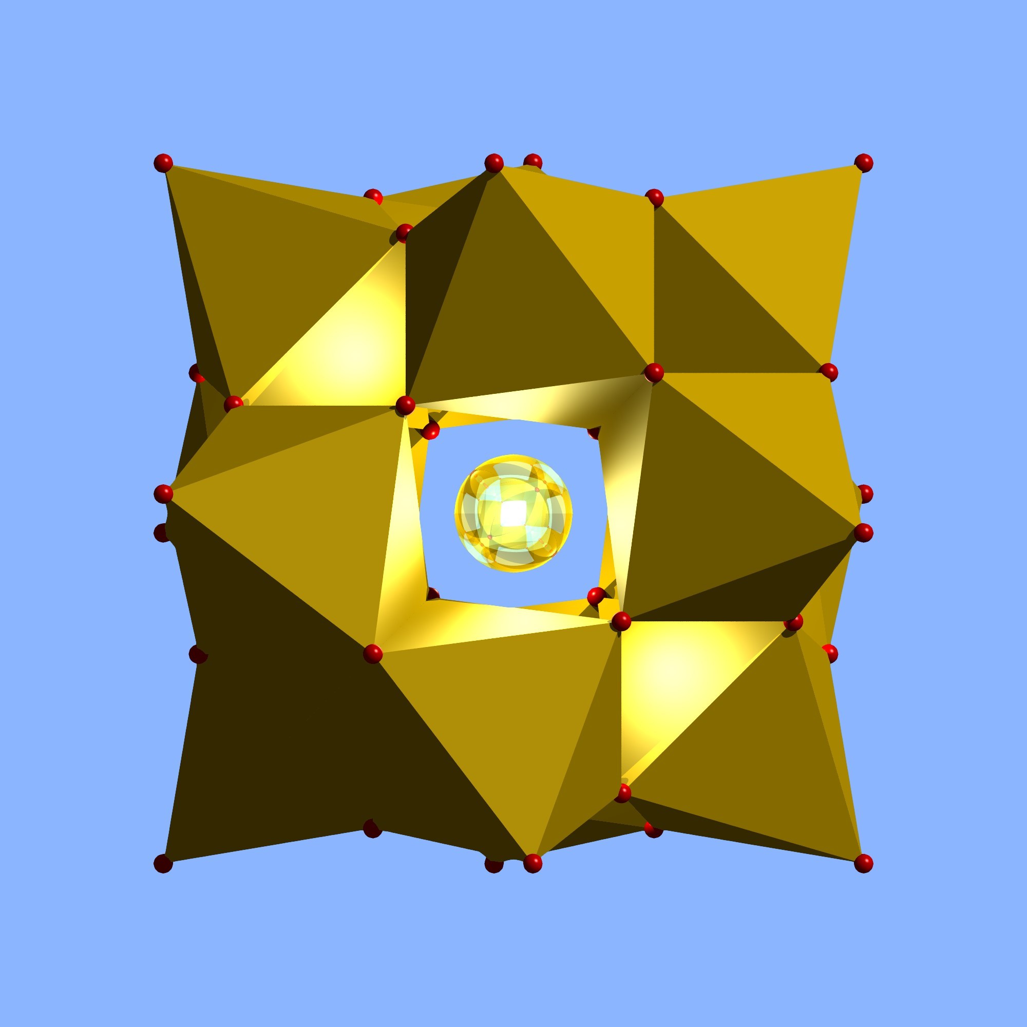
Diamond is our outstanding molecular and crystal structure visualization software. It integrates a multitude of functions, which overcome the work with crystal structure data - in research and education as well as for publications and presentations.

Diamond does not only draw nice pictures of molecular and crystal structures like most of its competitive programs do. It offers an extensive set of functions that let you easily model any arbitrary portion of a crystal structure from a basic set of structural parameters (cell, space group, atomic positions).
With its high data capacity, its wide range of functions beginning with the generation of molecules reaching up to the construction of rather complicated inorganic structural frameworks, Diamond is a comprehensive tool for both molecular and solid state chemists as well as for surface and material scientists.
Diamond, the well-known program for the visualization and exploration of crystal structures, has been improved again: Version 4 contains a lot of new features, in addition to many functions of earlier versions that have been enhanced significantly (like the definition of connectivity or the exploration of the structure).
New and Enhanced Features
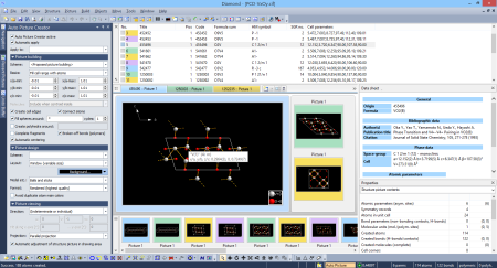
- "Auto Picture Creator" docking pane automatically applies changes in building options, picture design and viewing direction directly to the structure picture.
- "Grab mode": New mode for more intuitive rotation, shifting or zooming during exploration of a crystal or molecular structure.
- Intuitive structure exploration: preview neighboring atoms and molecules e.g. using the mouse wheel.
- Improved evaluation of bonding spheres (connectivity), including non-bonding contacts, improved handling of H-bonds, and determination of atom site environments using Dirichlet domains (Voronoi polyhedral).
- Easy application of user-defined design schemes.
- New options to create a packing diagram: cell range, sphere, slab, or slice of molecules.
- Creation of Voronoi polyhedral
- Improved functions to complete molecular fragments, generate symmetry-equivalent molecules, and search for non-bonding contacts.
- Expand or reduce coordination spheres around selected atoms, clusters of molecules, molecular fragments or polymers.
- Full screen view of structure picture.
- Access to the crystal structure database COD ("Crystallography Open Database").
- Improved searching of Diamond documents and structure files on your hard disk.
- Undo/Redo now with multiple steps together, assisted by thumbnail pictures of the previous conditions.
Diamond Function List
Please note: Functions that are new in version 4 - as compared with version 3.x - or have been enhanced significantly are emphasized. For functions that have been added to minor updates 4.x, have a look on the "New Functions..." link on the left side.
- 32 Bit MS Windows application with Multiple Document Interface (MDI), object-oriented menus, toolbars and local popup-menus. Allows 'simultaneous' handling of multiple structures.
Input and Output:
- Proprietary binary Diamond 3/4 Document format (extension .diamdoc) :
- Supports both crystal and molecular structures (i.e. with and without translational symmetry).
- Storage of multiple structure data sets in a document, each with:
- atomic parameters,
- cell parameters and space-group (optional),
- anisotropic displacement parameters,
- connection parameters (bonds, H-bonds, non-bonding contacts)
- Chemical and bibliographic data (author, reference, database origin, etc.).
- Supports multiple structure pictures for a structure data set. Saves your own built-up and designed frameworks of crystal structures.
- Compatible with Diamond 2 format (DSF).
- Number of atoms, bonds, polyhedral etc. limited only by RAM.
- Manual input or update of chemical, crystallographic, and bibliographic data.
- Automatic import from data formats:
- CRYSTIN download format created by ICSD or CRYSTMET
- Cambridge Structural Database FDAT format.
- Brookhaven Protein Data Bank format.
- SHELX-93 format.
- Crystallographic Information File (CIF).
- XYZ format (free format with Cartesian coordinates),
- SYBYL MOL and MOL2 format,
- Cerius2 (CSSR) format,
- MDL MOL format.
- Export of structure data to:
- CIF,
- SCHAKAL,
- XYZ format.
- POV-Ray assistant to create photo-realistic scenes with shadows, reflections, textures, background graphics, and more.
- Export of structure picture's 3D world to VRML.
- Export of structure picture's 2D graphics (for post-processing e.g. in a word processor or graphics application):
- as Windows metafile (WMF, vector-oriented),
- as bitmap (BMP; width, height and resolution user-defined),
- as GIF, JPG, or PNG file, e.g. to link with an HTML document.
- Cut, Copy, or Paste of data sets between documents (together with associated structure pictures). Enables creation of small "databases".
- Search for chemical, crystallographic, or bibliographic data:
- in files of selected types in selected directories,
- in "Crystallography Open Database" (COD), including (amongst others) AMCSD ("American Mineralogist Crystal Structure Database") as well as CIF files from the IUCr journals.
- Small database of most frequent (inorganic) structure types, e.g. to insert structure data or ready-defined structure pictures from.
- Configurable list of structure data sets in a document:
- as table ("structure list")
- or in collapsed form as "structure info bar".
- Color coding to differentiate structure data sets in a document with multiple structures.
- Thumbnail overview of structure pictures of a selected data set or of the whole document.
- Recent picture list with thumbnails of last edited pictures/documents.
- Data sheet for textual representation of structure data:
- in compact or comprehensive form
- or with customized selection and order of items.
- "Atom list": Hierarchical list of atoms in structure picture, groups by atom sites and/or groups or by molecules.
- Printing of selected datasets, data sheet, tables, or structure pictures. Textual copy of datasets via Windows clipboard for post-processing.
- Export of data sheet and tables as HTML.
Construction:
- Optional assistant that helps to create a structure picture from scratch or to modify a picture.
- "Auto Picture Creator" - available in a docking window side-by-side with the structure picture - to interactively change picture building as well as picture design and viewing direction.
- "Building schemes" (in Diamond 3 called "Auto-Builder") that create pictures automatically or according to a user-defined strategy. Useful when visualizing a lot of similar structures.
- Conversion between "crystal" and "molecular" structures, i.e. adding or removal of cell and symmetry information.
- Filling of unit cell, multiple cells, any cell range, or boxes or spheres around selected central atoms.
- Filling of user-defined rectangular areas within the screen.
- Filling of slabs along a plane (hkl or least-squares) or between a plane and the walls of the coordinate system.
- Selection of atoms to construct sublattices ("Filter").
- Creation (and discussion) of atomic environments, optionally from Dirichlet domains of the atom sites.
- Discussion of connectivity assisted by histograms showing the distribution of distances between selected atom types and from the bond parameters, together with automatic calculation and checking of distance ranges.
- Creation of bonds automatically, basing on connectivity, or manually by inserting bonds between two atoms each.
- Adding all atoms (and optionally bonds, H-bonds, contacts) of atomic parameter lists as well as from connection parameter lists.
- Generation of atoms from parameter list serving as initial atoms for building up complex frameworks.
- Completion of coordination spheres around selected atoms.
- "Pump up": Generation of multiple spheres around selected atoms and its reversal ("shrink").
- Automatic generation of molecules or completion of fragments which have e.g. been clipped at cell edges.
- Definition of molecular units (from atomic parameter list).
- Generation of molecules from molecular units at symmetry-equivalent positions.
- Search for molecules in the neighborhood of selected atoms or molecules.
- Creation of molecular packings (parallelepiped, sphere, slab, or layer).
- "Grow" and "cut”: Expansion and reduction of polymers or molecular fragments.
- Creation of "broken-off" bonds to signal infinitesimal chains, layers, or 3D-frameworks. Conversion between "broken-off" and normal bonds.
- Definition of H-bond and non-bonding contact connectivity. Creation of H-bonds and contacts.
- Expansion to neighboring atoms or molecules via H-bonds and/or contacts to build up molecule clusters and reversal ("reduce").
- Discussion of contact spheres and expansion or reduction of molecule clusters with the mouse wheel.
- Cut, copy and paste of structural parts between structure pictures:
- A fragment of a structure picture (or the whole picture) can be copied.
- The copied fragment can be pasted into a blank or another picture of the same data set.
- User-controlled dismantling of built-up frameworks.
- Multiple-step Undo and Redo function (with picture thumbnails) to enable safe experimentation with even high-complicated and unknown structural frameworks.
Visualization:
- "Design schemes" (a kind of style sheets) containing picture design and viewing settings for quick-and-easy application to other structure pictures.
- Layout modes:
- Regular/window,
- for printout, e.g. A4 page size with white background,
- for creation of a bitmap with given x and y dimension and a resolution in dpi.
- Variable zoom factor (enhances "Page view" mode of Diamond 2).
- Models, assigned globally or individually to single or groups of atoms (allows mixing of different models in one and the same picture):
- Ball-and-stick (regular),
- ellipsoid,
- space-filling,
- sticks or wires (depending on bond radius).
- Definition of views along special axes or toward special planes.
- Central or parallel projection, depth cueing, and stereo display.
- Photorealistic rendered models with user-defined light source and material properties (OpenGL).
- Variation of colors, styles and radii of atom groups and bonds. Individual design of each single atom is possible.
- Variation of atom and bond radii with mouse wheel.
- ORTEP-like atom styles (ellipses, octants) in both flat and rendering mode.
- Optionally fragmented and two-colored bonds.
- Labelling of atoms and bonds. User-defined text, can be placed at arbitrary position of picture.
- Generation of coordination polyhedral:
- Around central atoms of selected groups or around individually selected atoms,
- built up from selected ligand atoms,
- Optionally with transparent or hatched surfaces.
- Enhanced construction of coordination polyhedral:
- Defining corners and edges by selecting (clicking on) atoms and bonds, rsp.
- Removing of edges to increase triangles to higher polygons.
- Copy and Paste of polyhedron buildings between atoms of same site each.
- Definition of (transparent) lattice planes and (best) planes or lines through selected atoms.
- Adding of vectors to atoms to indicate e.g. a magnetic moment.
- Alternative color differentiation to visualize oxidation numbers, site occupation factors etc.
- Full screen view (with window frame, menu, toolbars hidden).
Animation:
- Movement of structure picture:
- Rotation along x-, y-, and/or z-axis,
- horizontal and/or vertical shift within drawing area,
- variation of enlargement factor (from Angstroems to centimeters),
- variation of camera distance (perspective impression).
- Controlled by:
- Mouse (the faster the mouse the faster the rotation etc.),
- keyboard (e.g. one degree rotation per keystroke),
- numerically (input through dialog).
- Optional "Spin" function, i.e. acceleration of movement.
- Continuous movement, which can be interrupted and continued.
- "Grab mode": Arcball rotation of an atom under the mouse cursor. Shifting with the right mouse button pressed. Changing the enlargement factor with the mouse wheel.
- Walk-through mode, enabling the camera/viewer to navigate through the structure picture.
- Recorder that helps to create video sequences, e.g. as AVI files.
- Creation of POV-Ray image sequences or videos from recorded pictures or from animations on a single structure picture.
Exploration:
- Neighborhood preview of atom under mouse cursor with radius of preview sphere variable with mouse wheel .
- Calculation of powder pattern:
- Variation of diffraction parameters:
- Radiation type: X-ray (laboratory, synchroton), neutron, electron,
- wavelength,
- LP correction,
- 2theta range,
- optional profile functions.
- Diffraction diagram (styles, colors and line weights can be configured).
- Table of reflection parameters with zoom in/zoom out and tracking through 2theta range.
- Calculation of distances and angles (incl. standard uncertainties):
- in a configurable table, for selected atom types and a sizeable distances range,
- around the atom(s) currently selected in structure picture.
- Graphical representation of distances as histogram with color-coded distances.
- Measuring of distances, angles, and torsion angles interactively (incl. standard uncertainties).
- Measuring of extended geometric features (incl. standard uncertainties):
- Angle between two planes (by hkl or (best) plane through 3 or more atoms),
- angle between two lines,
- angle between a normal of a plane and a line,
- distances of atoms from a plane or a line,
- centroid of a set of atoms,
- planarity or linearity of a set of atoms (distances of constituent atoms from plane/line).
- New Properties pane, displays information about:
- Contents of the structure picture (how many created atoms, bonds, polyhedral, etc.),
- the current "formula sum", that means the number of created atoms associated to atom groups,
- Info about the object that is selected in the structure picture or in the (optional) table above the properties pane, e.g. info about an atom of the parameter list,
- Table of the currently selected objects,
- Distances around the selected atom(s),
- Distances between the selected atoms,
- The center of the selected atoms (centroid),
- The planarity or linearity of the selected atoms and the deviations of the atoms from that plane or line, rsp.,
- Table of atoms assigned to the selected atom of parameter list or selected atom group,
- Table of bonds assigned to the selected bond group (i.e. atom group pair),
- Ligand, edges, and faces information of the selected polyhedral.
- Dirichlet domain of a selected atom site.
- Neighboring atoms and vertices of a selected Voronoi polyhedron.
System requirements
To install and run Diamond Version 4.0 or higher, you should have the following system requirements:
- Personal Computer with Microsoft Windows XP, Windows Vista, Windows 7, Windows 8/8.1 or Windows 10 operating system (note: does not run with Windows RT)
- Microsoft Internet Explorer 8 or higher
- Intel or AMD (or compatible) processor with "x86" instruction set
- 1 GB of RAM
- 3.8 GB of free disk space (6 GB during installation procedure, for unpacking of Crystallography Open Database)
- Graphics resolution of 1024 x 768 pixels with 32,768 colors ("High Color") or higher (1280 x 800 pixels or more recommended)
- DVD-ROM drive (for installation from DVD)
- Microsoft-compatible mouse

